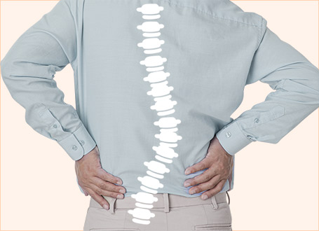
During the early days of our services, the standard diagnostic techniques were not very sophisticated. However, immense progress has been made in both diagnostic and surgical equipment tools in recent years. I have been a grateful beneficiary of this progress. 10 years ago I went through an artificial disc replacement surgery to treat severe lumbar and leg pain.
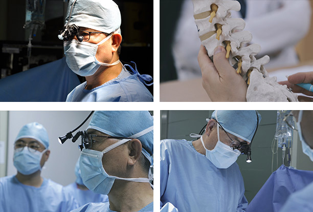
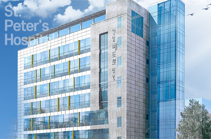
Inspired by my personal experience, Gangnam St. Peter’s Hospital has adopted the ‘root cause analysis’ approach.
Our methods result in 95% non-surgical and 5% surgical treatments for our patients. To date, we have performed upwards of 4,000 successful artificial disc replacement surgeries.
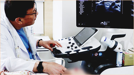
Today, St. Peter's Hospital Gangnam operates a total of 11 departments: Neurosurgical, Orthopedic, Rehabilitation Medicine, Internal Medicine, Cardiology, Nephrology (Dialysis Center), Thyroid Clinic, Urology, Obstetrics & Gynecology, and Physical Examination Center. Our skilled medical team of professionals operates a variety of state-of-the-art machinery (1x 3.0T MRI, 1x 1.5T MRI, 1x 640-channel MDCT high-performance CT scanner) to provide comprehensive, multidisciplinary care on par with any university hospital for our patients. We also offer 24-hour personal assistive care under our combined nursing & caregiving services.
Stroke, cerebral hemorrhage, cerebral infarction, Parkinson’s Disease, Alzheimer’s Disease
Disc Neutralization Therapy
This non-surgical therapy involves injecting drugs to neutralize acidified (pH 7.2 or lower) intervertebral discs. Disc neutralization therapy supplies nutrients to damaged discs and helps to recover elasticity and alleviate pain from impact. The flow of water, blood, and nutrients through discs is managed, helping damaged discs recover. Drugs are administered directly to the damaged areas, allowing for treatment of the root cause of pain. Disc neutralization is also an effective therapy against spinal stenosis, sciatic neuralgia, and chronic back pain.
Epidural Neuroplasty
After applying local anesthetic, live medical imaging device (C-arm) is used to identify the legion. Microcatheter (syringe 2mm in diameter) is inserted through the coccyx, and special preparation (local anesthetic, enzyme preparation, steroids, and hypertonic saline solution) is administered directly to the lesion. This rinses away inflammatory substances from the legion, dissolves scar tissues and adhered tissues, and reduces swelling of herniated discs and nerves, to promote healing of the disc. All while widening the spinal canal for uninterrupted passage of the nerves.
Collagen injection therapy
Instead of blocking and/or numbing pain for our patients, injecting ‘fibroblast collagen’ is similar to the already existing collagen in human bodies. The injection can promote disc regeneration and growth. This injection therapy not only removes spinal pain but also improves muscle strength.
Endoscopic discectomy
Minimal incision around 0.6cm is made on the skin. Damaged discs are removed selectively by endoscopic surgical tools. This minimally invasive method involves no bleeding or cutting of the bone, resulting in short recovery time. High-magnification lenses in the endoscope provide a large surgical field of view, allowing fine capillaries to be identified and avoided if needed, for safer and more accurate treatment. The procedure can be performed with local anesthesia and is safe for the elderly or diabetic patients with compromised immune functions, for whom general anesthesia may be dangerous.
Endoscopic Fusion
External filum terminale cutting addresses degenerative, not congenital symptoms, caused by the filum terminale of the peripheral spinal nerves pulling on the entire spine. The procedure is performed to treat syringomyelia, fibromyalgia, and restless legs syndrome, which occur more frequently in adults.
Microsurgery
This surgical method requires minimal cuts in the spine, preserving as
much normal tissue as possible while treating disc herniation or stenosis.
A microscope enters a tiny incision to magnify the lesion several tens of times. The procedure is low risk of damaging normal nerves, muscle, blood vessels, and cartilaginous plates in the vertebrae, providing high success rates with minimal risk.
Filum Terminale
This surgical procedure corrects meningocele in patients suffering from Arnold-Chiari Malformation. Cutting the filum terminale removes the root cause of tightness in the filum terminale of the peripheral spinal nerves. The method is performed through a perforation of the sacrum. At Gang nam St.Peter’s Hospital, external and internal filum terminale cutting procedures are performed according to individual patient’s conditions.
Lateral Lumbar Interbody Fusion (LLIF)
In the past, LLIF procedure was performed through anterior or posterior access. Today, improvements in understanding anatomical structures have allowed for artificial bone to be inserted in a bloodless procedure involving a minimal incision of about 2cm. Lateral access to the surgical site with minimal incision of skin and muscle tissue means fast post-op recovery periods, minimal damage, and aftereffects. The procedure can be performed safely on elderly patients as well.
Artificial Disc Replacement
In the past, lumbar fusion procedures were the norm. The purpose of these procedures was to prevent deformation or instability of the spine. However, these procedures limited freedom of movement in bending the neck or spine forward or backward. Artificial disc replacement was developed to address these disadvantages. Artificial disc replacement is a procedure where a severely damaged intervertebral disc is completely removed and replaced with an insertion of an artificial disc. This effective procedure immediately alleviates pain and minimizes degeneration of adjacent vertebrae while allowing for natural movement on par with a natural disc.
Microforaminotomy
This high-precision endoscopic spinal procedure is performed by a handful of surgeons in Korea. Microsurgery leaves an incision scar of around 3 cm. Palliative endoscopic surgery requires the use of 2 to 3 endoscopes at once. Microforaminotomy involves a single 0.5 to 1 cm fine incision followed by the insertion of a single endoscope containing a special camera and surgical equipment. The endoscope selectively removes damaged discs, muscles, ligaments, and bones.
Adult Stem Cell Therapy
Stem cells are called 'stem’ cells as they are the stems from which all types of cells are created. They have the ability to differentiate into any type of tissue found in the human body. St.Peter’s Hospital Gangnam offers adult stem cell therapies to repair damaged cartilage, regenerate damaged discs, and stop disc degeneration. These methods help to address the root cause of symptoms of pain.
Stroke, cerebral hemorrhage, cerebral infarction, Parkinson’s Disease, Alzheimer’s Disease
Chelation
Recurrence of cerebral infarction can be prevented using ‘chelation’, detoxifying injection therapy recognized by the American Heart Association. Acute cerebral infarction needs to be addressed with cerebrovascular treatment such as thrombectomy. Cerebral infarction is also a condition where treatment of aftereffects is crucial. The side effects of cerebral infarction can be remedied through rehabilitation therapy and stem cell therapy. Adult stem cell therapy.
Adult stem cell therapy
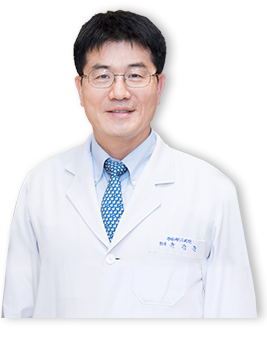
Stem cells are called 'stem’ cells as they are the stems from which all types of cells are created. They can differentiate into any type of tissue found in the human body. St. Peter’s Hospital Gangnam offers adult stem cell therapies to repair damaged cartilage, regenerate damaged discs, and stop disc degeneration. These methods help address the root cause of symptoms such as pain and pressure. Most intervertebral disc therapies focus on the surrounding ligaments, muscles, and nervous tissues affected by damaged discs. Chief of Medicine, Dr. Yoon Kangjun of Gangnam St.Peter’s Hospital has been treating neurological conditions (stroke, aftereffects of spinal cord injury, dementia, and Parkinson’s Disease, etc.) with adult stem cell therapy since 2001. Overcoming the limitations of conventional disc therapies, Dr.Yoon’s procedures inject adult stem cells into damaged discs to help regenerate the collagen fibers, elastic fibers, and proteins that make up the disc.
degenerative arthritis, cruciate ligament injury, meniscus injury, frozen shoulder, rotator cuff tear, carpal tunnel syndrome, hallux valgus
PDRN (DNA) injection
Cells similar to the actual cells found in the human body are injected into weak and degenerated tissues that cause chronic pain. This non-invasive therapy induces balanced regeneration through the natural mechanisms of the human body instead of artificial methods, and is effective in treating early spinal/joint conditions and post-surgical residual pain.
Cartilage Injection
To promote cartilage regeneration in damaged joints, injection therapy which contains a hyaluronic acid solution that contains the components of human cartilage and synovial fluid is introduced. Osteoarthritic patients are unable to naturally produce hyaluronic acid from their joints. Overtime, the amount of hyaluronic acid will decrease in their joints. This in turn causes reduced cartilage and synovial fluid elasticity, compromising the patient’s ability to protect their joints against external impact and aggravating the pain. Hyaluronic acid with high molecular weight is injected into the joints. This aids recovery to normal synovial fluid viscosity and elasticity. The joint will be able to move more smoothly, any impact on the joint will be better absorbed, joint functions will recover and pain will be reduced.
Extracorporeal Shockwave Therapy (ESWT)
This non-invasive therapy delivers 1,000 to 1,500 shockwaves to pain-causing legions, restoring blood flow in affected areas while stimulating the tissues and bone which reduce pain and restore function of the impacted area. The procedure usually takes around 30 minutes with no inpatient hospitalization. Patients can resume their normal day-to-day activities immediately. In most cases, the procedure is administered two to three times at 1-week intervals, depending on the patient’s condition. Patients are advised to avoid applying excessive force for 4 to 6 weeks following the procedure as they wait for the treatment to produce its effects.
Endoscopic Joint Surgery
Endoscopic joint surgery is a common procedure for arthritis, usually performed on knees and/or shoulders. A thin and long pencil-like apparatus around 5mm called an Arthroscope is inserted into the affected joint through a small incision in the skin. The arthroscope contains a small TV camera linked to a monitor, and the video feed from the camera is used to diagnose lesions and administer surgical treatment. The arthroscope is a long tube equipped with special lenses allowing for the structures inside a joint to be viewed from the outside. Also incorporated within the arthroscope are devices for illum inating and washing out the joint with water. The thickest arthroscope is 7.5mm across, meaning that the inside of a joint can be observed through an incision seldom exceeding 1cm. While the number of incisions depends on the procedure and the area of the joint the procedure is being performed on, on average 3 arthroscopes are used for viewing and irrigation. Technology such as the tiny surgical apparatuses have come a long way, endoscopic procedures have now replaced conventional knee joint surgeries. Currently, endoscopic procedures are used on joint cartilage surgery, synovial membrane surgery, fractured joint surgery and more. Arthroscopes were difficult to use when it first was invented, as the surgeon needed to look inside the tube with their bare eyes. The arthroscopes of the present produce a live video feed that can be viewed through a TV monitor. Photos and videos can be captured for data keeping. Endoscopic joint surgery can be performed with local or general anesthesia.
Artificial Joint Replacement Surgery
This procedure replaces damaged cartilage, which causes pain, with ergonomically shaped artificial cartilage. The process can be likened to grinding away dental caries and inlaying with gold. The advantage of artificial joint replacement surgery is that most patients experience no pain after the recovery period. Some patients complain of stinging or numbness in the joint following surgery. This is normal and occurs as the human body accepts the artificial joint. Reducing these symptoms requires consistent exercise, taking hospital-prescribed medicine for up to 3 months following the procedure, and frequent follow-ups. Patients having undergone knee joint replacement are urged to practice knee exercises to strengthen leg muscles. They are cautioned to avoid falling down to avoid damaging their artificial knee joint. If a patient does fall, they should visit a nearby hospital to have their knee checked. Intense exercise, walking for extended periods of time, and other strenuous activities that may strain the knees, should be avoided whenever possible. Artificial joints are made of metal and may trigger metal detectors during airport security screening or MRIs. Make sure to inform security staff of your artificial knee if any alarms are triggered, and also inform your dentist that you have undergone artificial joint replacement surgery.
Coronary artery, heart failure, arrhythmia, valvular heart disease, peripheral artery disease, hypertension, hyperlipidemia
Stent insertion
A stent is a small metal mesh made of special alloys which is inserted inside an artery. In the past, the balloon would be inflated inside the coronary artery to widen the artery. However, many patients undergoing this procedure would potentially suffer from another blockage a few months later. If a stent is inserted after widening the narrowed coronary artery, the artery remains widened. The stents used nowadays release drugs that prevent stenosis. Reducing stenosis recurrence rates after the procedure. Stents can be inserted using long tubes through the wrist or thighs for coronary angiography. Normally, the stents are installed during coronary angiograms when narrowing of the coronary artery has been discovered.
Coronary Artery Bypass Surgery
In cases involving severe stenosis, a coronary artery bypass procedure is the preferred alternative over stent insertion. As the name suggests, the bypass path is created around a blocked or narrowed portion of the coronary artery. Healthy sections of the artery are taken from the chest or legs to create the bypass. Unlike interventional procedures like stent insertion, coronary bypass surgery is performed under general anesthesia.
Chronic kidney disease
Blood Dialysis
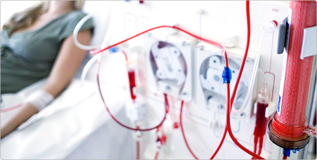
Blood dialysis is a procedure that uses an artificial kidney machine to filter the patient’s blood. Artificial kidney machine is made up of: blood pump, dialysate pump, and filter. The patient’s blood is pumped into the machine, where waste and excess moisture are filtered out. The newly cleaned blood is pumped back into the body. A single round of dialysis takes 4 hours. Patients typically require three rounds of dialysis a week. Dialysis is an effective way of regulating moisture and electrolyte levels in the blood. However, kidney disease complications such as arteriosclerosis and anemia, need separate drug therapy. St. Peter’s Hospital Gangnam operates a state-of-the-art artificial kidney center equipped with high-efficiency, high flow rate dialysis machines. Our kidney specialists work in conjunction with our other departments - cardiology, internal medicine, neurosurgery, orthopedics, urology, obstetrics & gynecology, thyroidology, and radiology - to provide patients with comprehensive, multidisciplinary medical attention. Gangnam St. Peter's Hospital is staffed round-the-clock by radiology experts performing hemodialysis access intervention and neurovascular intervention, to ensure faster and more accurate treatment. Maintaining and managing access is critical when performing blood dialysis.
St.Peter’s Hospital Gangnam operates a team of radiology experts at ready for hemodialysis access intervention procedures. The in-house team is immediately available in case of potential problems regarding access for blood dialysis. Percutaneous transluminal angioplasty (PTA) for arteriovenous fistulas, stenosis, blockages, as well as central venous catheter insertion, are available on site.
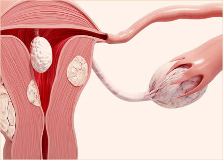
Uterine Myoma, Adenomyosis, Incontinence
HIFU
High-intensity ultrasonic waves are focused on tumor tissues in the body, the generated heat removes myoma. The uterus is preserved, enabling for
conception and childbirth.
Uterine Myoma Embolization
This procedure against uterine myoma selectively blocks off the uterine arteries on both sides of a myoma, inducing ischemia to reduce the size of the myoma.
Laparoscopic Surgery
An incision of 15~20 mm is made inside the belly button to insert the laparoscope and 2 surgical arms to remove the myomas.
Endocervical Surgery
A special scope equipped with special tools is inserted through the vagina. The myomas are removed with the scope offering a live view inside the uterus.
Hysterectomy
This surgical procedure removes the uterus altogether and involves prolonged hospitalization and long recovery times.
TOT therapy for incontinence
Transobturator tape or TOT therapy is a method of treatment where spinal anesthesia is administered and an ergonomically designed mesh tape for
incontinence treatment is used to secure the urethra in its normal position and provide support under abdominal pressure due to sudden coughing or
sneezing.
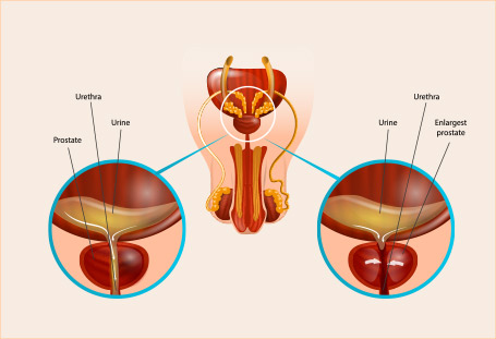
Prostatitis, prostatic hyperplasia, prostate cancer, urinary stones
HIFU
Prostate cancer HIFU is a new non-surgical method to treat prostate cancer. Focusing highly intense ultrasonic heat on prostate cancer cells causes necrosis of the target tumors. The dead cancer cells are dissolved naturally within the body and are excreted as waste.
Extracorporeal Shock Wave Lithotripsy (ESWL)
A method of treatment that uses a series of shockwaves to dissolve urinary stones
URS (ureteroscopy)
A flexible Olympus 4K ureteroscope is inserted through the urethra and bladder into the ureter to find, dissolve, and extract urinary stones.
Percutaneous Nephrolithotomy (PNL)
A method of treatment where an endoscope is inserted into the kidney through an incision in the back to separate and remove renal stones
Hypothyroidism, hyperthyroidism, thyroid nodule, thyroid cancer, benign breast nodule
BABA bilateral axillo-breast approach robotic thyroidectomy
BABA (bilateral axillo-breast approach) robotic thyroidectomy at Gangnam St.Peter’s Hospital, fine incisions (1 cm or smaller) are made around the armpits and the areola on the patient, which avoids incisions in the neck. Endoscopic surgical equipment is inserted into these incisions to remove thyroid cancer.
Transoral endoscopic thyroid surgery
NOTES (Natural Orifice Transluminal Endoscopic Surgery) is a scarless surgical procedure where endoscopes are inserted into 3 small incisions in the
mucosa between the lower lip and gums to remove cancer.
RFA radiofrequency ablation therapy
Radiofrequency ablation therapy is a method where a fine (1mm-thick) probe is inserted using real-time ultrasound into nodules to directly deliver
high-frequency heat at 100,000 to 500,000hz and cause necrosis. Radiofrequency waves are sent out from the probe into the surrounding tissue, which
causes the nearby cells to die. As these cells die, the immune system removes them, which causes an internal reaction and generally results in shrinkage
of the nodule.
Mammotome
The mammotome is a vacuum-like apparatus with a special 0.5cm needle which is inserted into the breast. Breast tissues are pulled toward the needle, and any breast tumors are removed. Mammotomes can be used not only for removal of benign breast nodules, but also for breast cancer biopsies.
At Gangnam St. Peter’s Hospital, rehabilitation specialists perform precise analysis of individual patients’ physical characteristics and exercise ability to administer rehabilitative therapy.
This program is called ‘1:1 Tailored Programs’.
Our exercise rehabilitation center offers a variety of therapeutic methods for improved patient satisfaction and therapeutic effect.
Rehabilitation therapies aim at addressing the root cause of pain and correcting the posture and body form to prevent recurrence.
Physiotherapy
Occupational therapy
Hyperhidrosis & bromhidrosis, hot flushes, Raynaud's syndrome, varicose veins, immune cancer treatment
What is Endoscopic Sympathectomy?
This is a minimally invasive spine surgery to treat hyperhidrosis. The procedure is effective at treating hyperhidrosis (sweaty hands, feet, armpits),
and facial blushing. The procedure is permanent and is administered frequently on patients where other treatments have failed.
Endoscopic Sympathectomy process
Microvascular Decompression (MVD)
Microvascular Decompression is a surgery to relieve abnormal compression of a cranial nerve such as hemifacial spasms.
This neurosurgical procedure targets the root cause of facial twitching, and has been in widespread use to treat facial twitching and facial paralysis for
over 30 years.